
What a control is doing in a flow experiment
A flow cytometry control is a sample run alongside your test sample to establish where a signal comes from. Controls tell you which events are genuinely positive for a marker, how much fluorescence spills between detectors, how much background your cells produce on their own, and whether the cytometer is performing the way it did last week.
Maecker and Trotter grouped controls into three classes in their 2006 tutorial, and the framework has held up: controls for instrument setup, controls for specificity and gating, and biological comparison controls.[1] Each answers a different question, and is critically important for a successful experiment.
Setup controls make the instrument behave consistently. Specificity controls tell you where the boundary between positive and negative sits. Biological comparison controls give you a reference population that should look a particular way, so you know whether the whole assay drifted.
The controls you'll actually set up
Unstained controls
Cells with no antibody, no viability dye, nothing. Their job is to show you autofluorescence, which varies enormously by cell type. Monocytes and macrophages are bright; lymphocytes are comparatively dim; anything with high flavin content will glow in the blue-excited channels, while NADH is excited by UV wavelengths instead.
An unstained control isn't a gating control and shouldn't be used as one. It shows you your floor, not your threshold.
Single-stained compensation controls
One fluorochrome, one detector, used to calculate spillover into every other detector. Get these wrong and every downstream population moves.
Three rules matter here. The control must be at least as bright as the sample, because dim compensation controls produce noisy spillover estimates. Positive and negative populations must be present in the same tube and must have the same autofluorescence. And the fluorochrome has to be the exact same conjugate, since tandem dyes vary between lots and between manufacturers.
Roederer's 2001 paper on compensation artifacts is still the clearest account of what goes wrong when these conditions aren't met, particularly the spreading error that no amount of correct compensation will remove.[2] Compensated data doesn't collapse neatly onto the autofluorescence level, and people spend a lot of time chasing that as if it were a settings problem.
Polystyrene beads are the usual answer for single-stain controls because they're cheap and consistent. They also scatter light nothing like a cell, carry no autofluorescence, and can shift the emission spectrum of a bound antibody relative to the same antibody on a cell surface. For most panels that's tolerable. For spectral unmixing, where the algorithm is fitting a full emission signature rather than a single spillover value, the mismatch matters more, which is why compensation and unmixing controls built to behave like cells solve a problem that beads structurally can't.
FMO controls
Fluorescence minus one: every fluorochrome in the panel except the one you're gating on. FMO controls show you how much signal bleeds into that channel from everything else, and they're the most defensible way to set a gate boundary on a continuous marker.
They're also expensive in sample and in time. A 12-color panel needs 12 FMO tubes if you run them properly, and most labs run them during panel development and then drop them for routine work. That's a reasonable compromise as long as everyone knows it's a compromise.
Isotype controls
An antibody of the same isotype, subclass, light chain and fluorochrome as your test antibody, raised against something the cells shouldn't express. The idea is to measure nonspecific binding.
The idea works less well than it looks. Hulspas and colleagues laid out the problems in their 2009 recommendations: matching isotype, subclass, light chain, fluorochrome and fluorochrome-to-protein ratio all at once is difficult, and an imperfectly matched isotype control tells you very little.[3] Isotype controls also can't account for spectral spillover, which means they should never be used to set compensation or to define the negative population in a multicolor panel.
They still have a place in specific circumstances, mostly when you're characterizing a new antibody clone or working with cell types prone to Fc receptor binding. Treat them as a diagnostic, not a routine gating control.
Viability controls
Dead cells bind antibody nonspecifically and will happily populate a gate you care about. A viability dye is standard practice; a viability control is the sample that tells you the dye is working and that your gate is in the right place.
Heat-killed or otherwise compromised cells give you a known-dead population. The problem is preparing them consistently, since heat-killing protocols vary and a batch prepared on Tuesday won't necessarily match Friday's. Labs running longitudinal studies or multi-site work need a viability reference that doesn't change, which is the argument for shelf-stable ViaComp viability controls over freshly prepared material.
Biological comparison controls
A sample that should look a particular way: unstimulated cells against stimulated, healthy donor material against patient material, a normal peripheral blood profile against an abnormal one. Maecker and Trotter argued these often give a better positive-negative boundary than isotype or FMO controls, because they carry the same biology as the test sample.[1]
They're also the hardest controls to standardize, and that difficulty is the whole reason this category of problem exists.
Control selection table
| Question you're asking | Control to run | What it won't tell you |
|---|---|---|
| Is the cytometer performing consistently? | Calibration particles, daily QC beads | Anything about your staining |
| How much do my cells glow on their own? | Unstained sample | Where to set a gate |
| How much signal spills between detectors? | Single-stained compensation controls | Whether staining is specific |
| Where does positive start on a continuous marker? | FMO control | Nonspecific antibody binding |
| Is this antibody binding specifically? | Isotype or isoclonic control | Spillover, or the negative boundary in a multicolor panel |
| Are dead cells contaminating my gate? | Viability dye plus a known-dead reference | Antibody specificity |
| Did the whole assay drift since last run? | Biological or reference control | Which step drifted |
| Do two instruments give the same answer? | Scatter and fluorescence reference material run on both | Whether either is correct in absolute terms |
| How many antigens per cell? | Quantification standards with assigned ERF values | Anything about the antigen's function |
Where control materials break down
Two failure modes account for most of the reproducibility problems labs run into, and neither is a technique error.
Donor material varies. Peripheral blood mononuclear cells from different donors express markers at different densities, carry different autofluorescence, and behave differently after freeze-thaw. Use donor cells as your reference standard and your reference standard changes every time you change donor. For a single experiment that's survivable; for a study running eighteen months across four sites, it isn't.
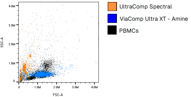
Beads aren't cells. Polystyrene microspheres are wonderfully consistent, which is exactly why they're the default for compensation. They also sit in a completely different place on a scatter plot, carry none of the autofluorescence a real cell carries, and can't stand in for a cell when the question involves scatter, viability or antigen density. Anyone who's tried to set a lymphocyte gate using a bead control knows this already.
Cell mimics exist to sit between those two failures: engineered particles with cell-like scatter and controllable fluorescence, manufactured to a specification rather than harvested from a donor. Scatter-matched standardization and immunophenotyping controls both depend on that combination, and it's worth understanding how cell mimics are built before deciding whether they solve your particular problem.
What the standards say
CLSI published H62, Validation of Assays Performed by Flow Cytometry, in October 2021.[4] It's the closest thing the field has to formal guidance on validating cell-based flow assays, covering instrument qualification, assay optimization, analytical method validation and post-examination practice.
One point in H62 is worth sitting with. Validating a flow cytometry assay is harder than validating a biochemical one partly because flow data doesn't come from a calibration curve, and partly because true reference standards are lacking for most measurands. That's a candid admission from a consensus document, and it explains why control material selection carries so much weight in this field: there's frequently nothing else to anchor to.
Monaghan and colleagues built on H62 in 2025 with recommendations covering what happens when a validated method gets modified, including which validation parameters need re-evaluating for each type of change.[5] Swapping a control material counts as a modification, so this is worth reading before you change anything in a validated assay.
Choosing controls for a regulated assay
Research panels and regulated assays have different constraints, and the difference is mostly about time rather than rigor.
For research work, choose controls that answer the question in front of you and accept that you'll rebuild them when the panel changes. FMO controls during development, single-stains matched to your conjugates, an unstained tube every run.
For anything heading toward a regulatory filing, the constraint becomes availability. A control material you can't source in the same specification in three years is a control material that will force a partial revalidation halfway through your study. Lot-to-lot consistency, documented specifications, and a supplier who can commit to continuity matter more than any single performance characteristic. This is where donor-derived reference material tends to fail, not because it performs badly but because it runs out.
Custom control material is worth considering when your marker of interest doesn't have a commercial equivalent, which is common in cell and gene therapy work. Custom biomarker cell mimics can be built to a defined antigen density, which also opens up quantitative antigen density measurements that aren't possible with a qualitative control.
Common questions
- What are the main types of flow cytometry controls?
- Unstained controls, single-stained compensation controls, FMO controls, isotype controls, viability controls, and biological or reference controls. They divide into three functional classes: instrument setup, specificity and gating, and biological comparison.
- Do I still need isotype controls?
- Rarely, and not for setting gates in a multicolor panel. They can't account for spectral spillover and they're difficult to match properly. FMO controls or well-chosen biological controls are better for defining a positive boundary.
- Can I use compensation beads for everything?
- No. Beads work well for calculating spillover when the conjugate matches your sample exactly. They don't reproduce cellular scatter or autofluorescence, so they can't serve as viability controls, scatter references, or gating controls.
- What's the difference between a control and a standard?
- A control tells you whether the assay behaved as expected on a given run. A standard carries an assigned value you calibrate against, such as antigen binding capacity for quantitative work. Most flow assays have controls; comparatively few have true standards, which is one of the gaps CLSI H62 identifies.
- How many controls does a multicolor panel need?
- At minimum, one unstained sample, one single-stained control per fluorochrome, and a viability control. FMO controls are added per marker during panel development. A 12-color panel run properly during development can involve 25 or more control tubes, which is why most labs reduce the set once the panel is locked.
References
- 1.Maecker HT, Trotter J. Flow cytometry controls, instrument setup, and the determination of positivity. Cytometry Part A. 2006;69A(9):1037-1042. doi:10.1002/cyto.a.20333
- 2.Roederer M. Spectral compensation for flow cytometry: visualization artifacts, limitations, and caveats. Cytometry. 2001;45(3):194-205. doi:10.1002/1097-0320(20011101)45:3<194::AID-CYTO1163>3.0.CO;2-C
- 3.Hulspas R, O'Gorman MRG, Wood BL, Gratama JW, Sutherland DR. Considerations for the control of background fluorescence in clinical flow cytometry. Cytometry Part B: Clinical Cytometry. 2009;76B(6):355-364. doi:10.1002/cyto.b.20485
- 4.Clinical and Laboratory Standards Institute. H62: Validation of Assays Performed by Flow Cytometry. 1st ed. Wayne, PA: CLSI; 2021.
- 5.Monaghan et al. Flow cytometry assay modifications: recommendations for method validation based on CLSI H62 guidelines. Cytometry Part B: Clinical Cytometry. 2025;108(3):252-266. doi:10.1002/cyto.b.22202