Understanding where T cells sit along the activation-exhaustion spectrum is central to modern immunology. Whether building broad immuno‑oncology panels, characterizing CAR T‑cell products, or evaluating tumor‑infiltrating lymphocyte (TIL) potency, researchers rely on the combined readout provided by multicolor flow cytometry to interpret functional state with simultaneous identification of a range of phenotypes of T cells within the same sample.
Generating robust biological controls has been a longstanding challenge in flow cytometry, and this challenge is amplified for checkpointassociated markers. Early activation markers such as CD69 and CD25 are transient and can shift within hours depending on stimulation strength, culture conditions, and donor biology. Exhaustion markers including PD1, TIM3, LAG3, CTLA4, and TIGIT are typically dim, rare, and highly contextdependent, often requiring prolonged or repeated stimulation to induce stable expression. As a result, biological controls for activation and exhaustion demand substantial preparation, are difficult to reproduce consistently, and frequently yield populations that are too small or too variable to serve as reliable references.
Large standardization efforts such as the EuroFlow Consortium have shown that even basic immunophenotyping requires strict harmonization of instrument settings, reagent handling, and gating strategies to achieve reproducible results across operators and sites (Kalina et al., 2012). More recent work developing standardized PBMC immunophenotyping workflows which include activation and exhaustion markers highlights their sensitivity, concluding that careful gating controls are required for repeatable assay performance even for welltrained operators (Kiel et al., 2026).
Slingshot Biosciences’ T Cell Activation/Exhaustion Mimic™ was designed to remove this burden by providing stable, reproducible synthetic populations that model distinct stages of Tcell activation and exhaustion. These controls support consistent panel design and gating strategy optimization for CD8+ activated and exhausted Tcell populations, eliminating the need to generate separate biological controls.
Defined, stable populations for CD8+ T-cell functional states
The T Cell Activation/Exhaustion Mimic™ contains defined populations that represent biologically meaningful checkpoints in CD8+ Tcell progression (Figure 1).
- Pan CD8+ T cells CD45+, CD3+, CD8+ A baseline phenotype with no activation or exhaustion marker expression.
- Early Activated T cells CD45+ CD3+ CD8+ CD25+ CD69+ PD-1- Captures the short-lived early activation window where CD69 and CD25 are co-expressed before inhibitory receptors appear.
- Late Activated T cells CD45+ CD3+ CD8+ CD25+ CD69- PD-1+ Represents sustained activation with early checkpoint engagement.
- Exhausted T cells CD45+ CD3+ CD8+ CD25- CD69- PD-1+ This population can be further resolved for TIM-3, LAG-3, CTLA-4, and TIGIT, providing reliable access to inhibitory receptors that are rarely co-expressed in healthy donor PBMCs.
Each vial also includes a synthetic “dead” population compatible with both amine‑reactive and DNA‑intercalating viability dyes. This provides a consistent viability reference without relying on damaged or heat‑killed biological samples, which can vary significantly from run to run.

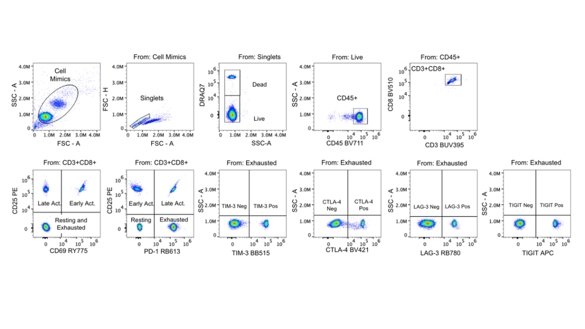
Figure 1. T Cell Activation/Exhaustion Mimic™ example gating strategy. Here, a DNAintercalating viability dye(DRAQ7) has been used to define a live gate. Each population is present at predefined concentrations within eachvial, providing clear gating guidance that can be applied to experimental samples.
How synthetic controls can improve activation/exhaustion assays
Activation markers without additional biologics handling
Mimic controls provide activation markers such as CD69 and CD25 without the need to stimulate PBMCs. This allows for greater consistency in the abundance and intensity of activation markers and reduces the handson work required to generate activation controls.
Exhaustion markers without chronic stimulation models
Exhaustion markers including PD‑1, TIM‑3, LAG‑3, CTLA‑4, and TIGIT are present in defined exhausted populations, supporting template development, panel optimization, and longitudinal studies without relying on chronic stimulation models.
Removing doubt with stable, reliable controls
Because the mimics are synthetic, they do not drift over time or vary by donor. This stability makes them well‑suited for multi‑site assay harmonization, longitudinal immune monitoring, system suitability testing, and QC workflows. They also align with best‑practice immunophenotyping guidance, which emphasizes the importance of well‑defined controls when interpreting dynamic immune markers.
Built on the same reliable technology as other Slingshot products, T Cell Activation/Exhaustion Mimic™ offers robust and stable lot‑to‑lot performance. Each vial provides multi‑year stability and is produced in large, uniform manufacturing lots. A peer‑reviewed analytical validation of the related TBNK Mimic™ product reported ≤5% CV for intra‑ and inter‑assay precision, linearity of R>0.998, and <5% drift under accelerated stability testing (Gunturu et al., 2026).
A practical foundation for activation/exhaustion panels
The T Cell Activation/Exhaustion Mimic™ provides a reliable, reproducible foundation for studying some of the most biologically complex and clinically relevant Tcell phenotypes. These defined synthetic populations enable consistent gating and panel optimization across activation and exhaustion states, eliminating the variability and extensive preparation required for biological controls.
References and links
Kalina T, FloresMontero J, van der Velden VHJ, et al. EuroFlow standardization of flow cytometer instrument settings and immunophenotyping protocols. Leukemia. 2012;26(9):19862010. doi:10.1038/leu.2012.122
Kiel MJ, Biswas S, Martinez A, et al. A standardized singletube 17color spectral flow cytometry workflow for integrated immunophenotyping of human PBMCs and mixed coculture systems. Front Immunol. 2026;17:1799028. doi:10.3389/fimmu.2026.1799028
Gunturu SP, Biswas S, Martinez A, et al. Beyond PBMCs: polymer‑based cell mimics for robust TBNK immunophenotyping assay validation. Cytometry A. 2026;109(4):290‑306. doi:10.1002/cyto.a.70031
T Cell Activation/Exhaustion Mimic™ — Immunophenotyping Controls
Stay Updated
Subscribe to receive product updates, new resources, webinars, and application notes.