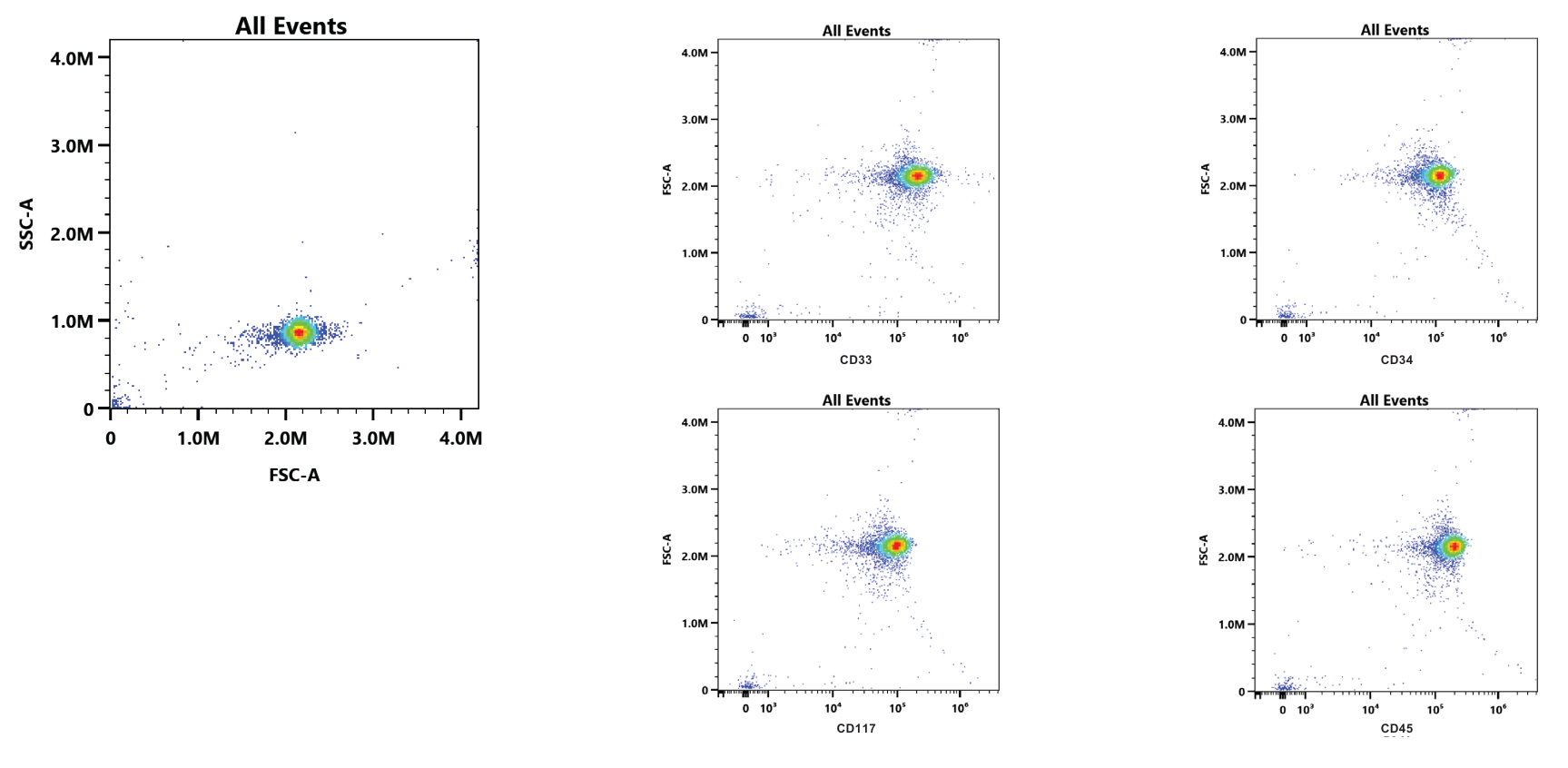
- 4 markersCD34, CD117, CD45 and CD33 on one cell mimic
- ~10⁵ MFITarget expression for every biomarker
- Up to 1 yearStability when properly stored
Project overview
Biological reference materials are critical in supporting the basis of comparative analysis in clinical testing. Often crucial key decisions, such as a clinical diagnosis or determining a treatment strategy for a patient, can be decided by the outcome of such comparative testing. Timeliness of sourcing, innate variability of donor material, and overall sample integrity can limit the outcomes of clinical trials and negatively impact disease diagnosis.
A prominent global pharmaceutical company was addressing the challenge of an appropriate control for acute myeloid leukemia (AML) by using the Kasumi-3 cell line as diagnostic reference QC material. However, there were issues associated with the low expression of key markers on Kasumi-3 cells. The company reached out to Slingshot Biosciences to develop a synthetic Kasumi-3 AML cell line expressing CD34, CD117, CD45, and CD33 to mimic the AML myeloid lineage at a target Mean Fluorescence Intensity (MFI) of ~10⁵ for all biomarkers. The senior scientist stated, “We needed a material that could act as a staining control to monitor inter-run assay performance for clinical sample analysis performed from fresh whole blood samples collected on study - ideally, a material that is readily available that could be easily prepared.”
After a successful partnership, the company determined that the performance of the Slingshot Biosciences synthetic Kasumi-3 cell mimic is similar to a staining control of Kasumi-3 cells.
Customer AML control challenges
- High variation in antigenic expression between patient samples and healthy donors makes it difficult to classify or diagnose hematological malignancies
- Significant costs associated with cell line maintenance, including infrastructure costs, reagent costs, and costs to source primary disease cells
- Impact of cell line maintenance on time to commercialization and assay development as resources and time are needed for bridging studies to address lot-to-lot variability
- Short shelf-life and usability of a sourced batch of cell lines in terms of limited useful passages from a single vial as well as issues of low viability or cell loss
Solution
Slingshot Biosciences’ innovative technology allows for the generation of synthetic cell lines that mimic any disease state. FlowCytes® are shelf-stable at room temperature, non-biohazardous synthetic cells that resemble the optical properties of human blood populations for lymphocytes, monocytes, and granulocytes. These FlowCytes can be customized to express any desired marker and antigen density.
Using this technology, the Slingshot team engineered a synthetic cell line expressing all markers requested (CD34, CD117, CD45 and CD33) at a target MFI of ~10⁵ (Figure 1). These markers have been identified as the most important markers for identification of myeloid blasts, a diagnostic feature of AML.¹
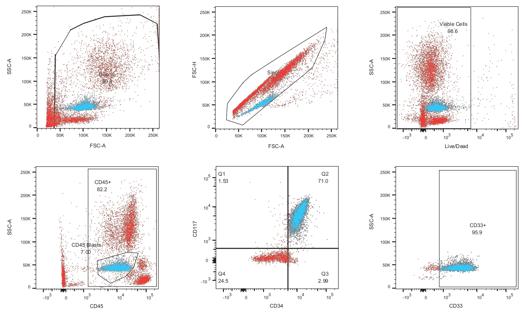
When the Slingshot synthetic AML cells were spiked with whole blood from a normal healthy donor and stained with relevant antibodies, the back-gating analysis showed similar light scatter profile and antigen expression compared to leukocytes (Figure 2). The CD45 vs SSC plot is a known strategy for identifying the blast gate, which the Slingshot team was able to mimic in the engineered process control (Figure 2, bottom left).
Outcome
The synthetic AML mimic from Slingshot serves as a more reliable control that accurately mimics the disease state. Use of these synthetic cell lines is a significant contributing factor to the accuracy of AML diagnosis, and can also help determine appropriate antibody titration levels.
The customer plans to utilize these cells for quality control purposes to develop future assays. These synthetic cells were purchased in bulk and, because of their long shelf life and stability, will ultimately lead to faster turnaround times for assay development and improve long-term assay stability.
In addition, use of the Slingshot AML cell mimic eliminates the need for significant cell culture infrastructure as these are non-biohazardous and stable for up to a year when properly stored. The enhanced lot-to-lot consistency of the synthetic AML cell mimic also dramatically reduces the resources required for bridging studies to address biologically derived controls that drift.
Most labs are currently forced to coordinate with a tissue culture facility to confirm whether cells are available for testing on a particular day, with sufficient quality metrics compared to the previous set of experiments. More often than not, the cells that are available are not from the same batch, leading to extensive quality control studies to maintain assay consistency. Slingshot’s vision is for customers to be able to readily pull a vial of disease control synthetic cells and, in some cases, add them to healthy donor blood, creating a customized biomarker profile for virtually any application.
The Slingshot advantage
- Level-up with truly cell-like performance
- Our synthetic cells match the optical, fluorescence, and biochemical features of cells
- Customizable to any cell population
- Precisely control the level of key biomarkers
References
- Wood, Brent L et al. “2006 Bethesda International Consensus recommendations on the immunophenotypic analysis of hematolymphoid neoplasia by flow cytometry: optimal reagents and reporting for the flow cytometric diagnosis of hematopoietic neoplasia.” Cytometry. Part B, Clinical cytometry vol. 72 Suppl 1 (2007): S14-22. doi:10.1002/cyto.b.20363
“Slingshot has developed an innovative product that could have implications for a variety of applications related to clinical flow cytometry.”
Stay Updated
Subscribe to receive product updates, new resources, webinars, and application notes.
Get the full case study
Download the complete write-up, with methods, figures and references, to share with your team.
Download PDF