Introduction
This is an important aspect of flow cytometry experimental setup for multiple reasons. In addition to emitting higher autofluorescence, dead cells also tend to experience much higher levels of non-specific binding. Both factors can skew results towards an elevated population of false positives for one or multiple markers. This can be especially harmful to experiments that hope to evaluate a population that is weakly positive for a marker of interest. In research, eliminating dead cells from the analysis is a precursor to obtaining accurate data and ensuring consistency of results across multiple experiments.
Viability staining is also an essential component of flow cytometry for therapeutic applications, such as the use of Fluorescence-Activated Cell Sorting (FACS) to transplant a subset of donor cells into a patient. For such treatments, proper live/dead staining is vital to ensure that patients receive only the healthy percentage of the sample during transplant, rather than an immunogenic dose of dying or damaged cells.
As such, gating to eliminate the dead cell population should occur before subsequent gates and analyses are established. This necessitates a viability control that will clearly delineate where populations of live and dead cells will appear.
Viability controls are generally a mixture of live and dead cells stained with a differentiating dye. These dyes generally fall into one of two broad categories: DNA-intercalating and primary amine (protein) stains. While these two types of dyes naturally have different targets, both rely on the increased cytoplasmic and nuclear membrane permeability of dead cells, and thus brighter labeling of this population.
As hinted previously, most current protocols rely on killing a portion of the experimenters’ sample and mixing this with live cells to create a viability control. Not only is this time-consuming and potentially wasteful (especially if the researcher is working with a rare or difficult to obtain cell sample), but it often relies on harsh chemicals to facilitate cell death. Furthermore, viability controls prepared in this way may still fail to achieve the unambiguous separation of live and dead cell populations that is needed for optimal gating.
In response to these obstacles, ViaComp® was developed. This unique viability control represents the first of its class. This reagent contains conveniently pre-mixed live and dead synthetic cells that are ready to be labeled with the DNA-intercalating or primary amine dye of your choice.
In this application note, we illustrate the use of ViaComp® by Slingshot Biosciences. Using the Cytek® Northern Lights 5-laser cytometer and their SpectroFlow® software, we assessed the performance of ViaComp® viability controls in conjunction with both DNA-intercalating and primary amine dyes. Altogether, the results presented here illustrate the superior ease-of-use and the dependable performance of ViaComp® compared to older, alternative viability control reagents and methods.
Results
Viability staining is an indispensable part of flow cytometry experimental setup. This step, followed by the subsequent elimination of dead cells from an analysis reduces false-positive signals and maintains the consistency of results across experiments. In therapeutic applications, accurate live/dead staining assures that the sorted cells being re-administered to a patient are competent.
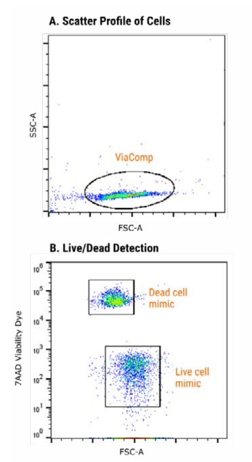
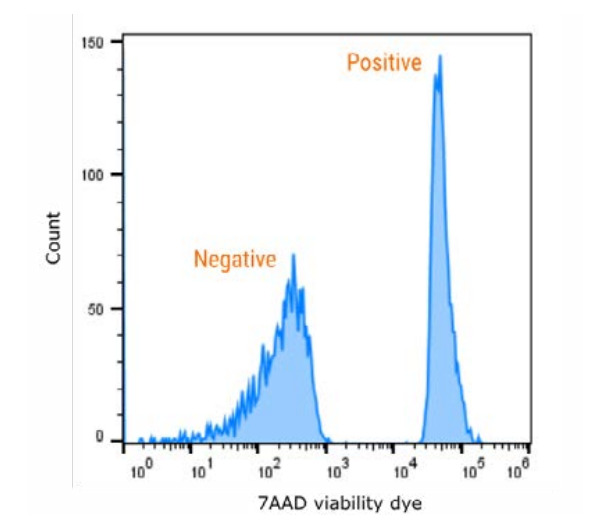
DNA intercalating dyes are one of two frequently used markers of live and dead cell populations. DNA-intercalating dyes, such as 7-AAD, will stain only non-viable cells by permeating the compromised plasma and nuclear membranes of these dead cells and intercalating, or inserting non-covalently, between base pairs of DNA. These dyes will brightly stain dead cells while viable cells remain unlabeled. As demonstrated by Figures 1 and 2, ViaComp® conveniently works with an array of the most popular DNA-intercalating dyes, including 7-AAD.
Performance of ViaComp® in achieving clean and defined separation of populations is largely unmatched; setting up accurate gates during data analysis becomes entirely seamless when using these live and dead cell mimics. Furthermore, as shown by Figure 2, the mean fluorescence intensity (MFI) achieved by the positive, 7AAD-containing cell mimics is high, providing an additional layer of dependability.
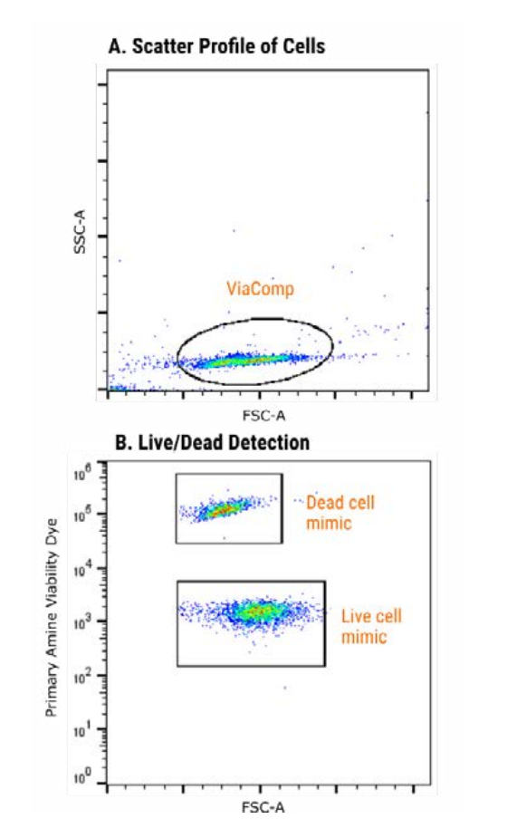
Primary amine dyes represent another method of differentiating between live and dead cell populations. PA dyes depend on a similar principle of membrane permeability but instead ubiquitously target proteins. In the case of live cells with intact membranes, this constitutes only the surface proteins, while in dead cells, both the surface and interior proteins will be labeled. Live/dead staining with PA dyes thus relies on the comparatively much brighter signal of dead cells. A notable advantage of this category of label is its effectiveness when used in conjunction with fixed cell samples.
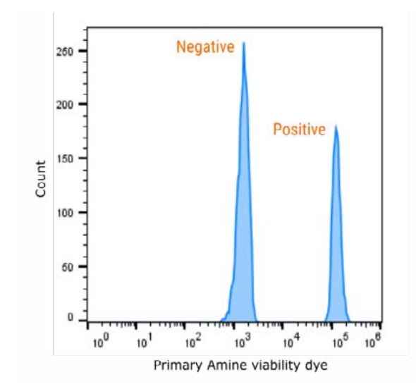
ViaComp® similarly demonstrates high performance when paired with commonly used primary amine dyes. Figure 3 demonstrates the clear separation of live and dead populations when ViaComp® is used in conjunction with a popular PA dye that excites at 488nm. Again, as shown in Figure 4, the ViaComp® positive stained cell mimics reach an intensely bright MFI, and the positive and negative populations are widely separated and well-defined.
Conclusion
Viability staining is important both in experimental research and therapeutic applications of flow cytometry. Currently, common viability staining methods require researchers to kill a portion of their cells in an added step that is wasteful, time-consuming, and often still does not achieve the desired standard of control. ViaComp® is a one-of-a-kind viability control containing conveniently pre-mixed live and dead synthetic cell mimics. ViaComp® dead cell mimics are DNA-containing, making this reagent a full process control for DNA-intercalating live/dead labeling, and this reagent works beautifully with a wide array of both DNA and primary amine dyes. The results detailed in this application note demonstrate the seamless usability and paradigm-shifting performance of ViaComp® in delineating live and dead population gates as well as in achieving an overall bright and reliable signal. In short, ViaComp® is a uniquely convenient, clean, and dependable viability control that can be readily integrated into your existing flow cytometry workflows.
References
- Bonetti, Maria Ida, Laura Pieri, Lola Domenici, Serena Urbani, Giovanni Romano, Alessandra Aldinucci, Clara Ballerini, et al. “Dendritic Cells with Lymphocyte-Stimulating Activity Differentiate from Human CD133 Positive Precursors.” Blood 117, no. 15 (April 14, 2011): 3983–95. https://doi.org/10.1182/blood-2010-08-299735.
- Burns, Jeremy Carlos, Bunny Cotleur, Dirk M Walther, Bekim Bajrami, Stephen J Rubino, Ru Wei, Nathalie Franchimont, Susan L Cotman, Richard M Ransohoff, and Michael Mingueneau. “Differential Accumulation of Storage Bodies with Aging Defines Discrete Subsets of Microglia in the Healthy Brain.” ELife 9 (June 24, 2020): e57495. https://doi.org/10.7554/eLife.57495.
- Davies D. “Viability Dyes for Flow Cytometry: It’s Not Just a Matter of Life and Death.” BiteSizeBio (January 6, 2015) https://bitesizebio.com/22353/viability-dyes-for-flow-cytometry-its-not-just-a-matter-of-life-and-death/
- Feng J, van der Zwaag M, Stokman MA, van Os R, Coppes RP. Isolation and characterization of human salivary gland cells for stem cell transplantation to reduce radiation-induced hyposalivation. Radiotherapy and Oncology. 2009;92(3):466-471. doi:10.1016/j.radonc.2009.06.023
- Kozlova, Anastasiia A., Roman A. Verkhovskii, Alexey V. Ermakov, and Daniil N. Bratashov. “Changes in Autofluorescence Level of Live and Dead Cells for Mouse Cell Lines.” Journal of Fluorescence 30, no. 6 (December 2020): 1483–89. https://doi.org/10.1007/s10895-020-02611-1.
- Kummrow, A., M. Frankowski, N. Bock, C. Werner, T. Dziekan, and J. Neukammer. “Quantitative Assessment of Cell Viability Based on Flow Cytometry and Microscopy.” Cytometry Part A 83A, no. 2 (February 2013): 197–204. https://doi.org/10.1002/cyto.a.22213.
- Perfetto, Stephen P., Pratip K. Chattopadhyay, Laurie Lamoreaux, Richard Nguyen, David Ambrozak, Richard A. Koup, and Mario Roederer. “Amine-Reactive Dyes for Dead Cell Discrimination in Fixed Samples.” Current Protocols in Cytometry 53, no. 1 (July 2010). https://doi.org/10.1002/0471142956.cy0934s53.
- Salas-Massó, Nuria, Quyen Than Linh, Wai Hoe Chin, Anders Wolff, Karl B. Andree, M. Dolors Furones, María José Figueras, and Dang Duong Bang. “The Use of a DNA-Intercalating Dye for Quantitative Detection of Viable Arcobacter Spp. Cells (v-QPCR) in Shellfish.” Frontiers in Microbiology 10 (February 28, 2019): 368. https://doi.org/10.3389/fmicb.2019.00368.
Featured product
ViaComp®
slingshotbio.com/products/viacompSSB-07-A
 View all product info
For Research Use Only. Not for use in diagnostic or therapeutic procedures.
Stay Updated
Subscribe to receive product updates, new resources, webinars, and application notes.
Get the full application note
Download the complete note as a PDF, with the figures and reference, to share with your team.
Download PDF