- 18.97complexity index with ViaComp® Ultra™ XT – Amine, vs 22.98 for polystyrene beads
- 36-colorspectral panel adapted from OMIP-112, reduced from 42 parameters
- 15 of 36reference controls first substituted with SpectraComp® and ViaComp® cell mimics
- 1 controlsingle, universal unmixing control for both viability and antibody reference spectra
Abstract
Panel development and optimization in flow cytometry is a standard part of developing high complexity multi-color panels. Ideal detection and measurement of chosen cellular markers in spectral flow cytometry requires single stained reference controls that can display the authentic fluorophore spectra and that are highly similar to the autofluorescence signature of biological cell samples. In this study, a 36-color immunophenotyping panel was initially developed using polystyrene beads, however switching select fluorophores to Slingshot Bio’s cell mimic SpectraComp® and ViaComp® compensation/spectral unmixing controls improved unmixing for problematic channels. To get the best unmixing results for this panel with the most straightforward workflow, ViaComp Ultra XT™ – Amine was used as a single, universal unmixing control for both viability and antibody reference spectra.
Introduction
Development of high-parameter panels for spectral flow cytometry can be a time-intensive, iterative process. Resolution of the true spectral signatures is critical for accurate detection of biological antigen expression patterns. However, the complex spectral composition of these samples are difficult to untangle. As more fluorophores are added to the panel, the rate of overlapping emission spectra increases, which makes detecting the true fluorophore signal through the noise a challenge. In addition, cells have their own auto-fluorescent signatures driven by intrinsic cellular components, such as aromatic proteins and flavins.¹ Deconvolution of these overlapping signals is essential for analysis.
Spectral unmixing is the process used to mathematically calculate fluorochrome abundances from raw detector values measured on a flow cytometer.² Accurate unmixing requires a set of single-stained reference controls that accurately represent the emission spectrum of each fluorophore in the antibody panel used for flow cytometry analysis. If reference fluorophores are mismatched with antibody staining on cells, unmixing errors can result.³ Therefore, the control selection is a critical step in experimental development.
Cells stained with individual fluorochrome-conjugated antibodies or viability dyes can be used as reference controls for spectral unmixing. The benefit to using cells is that they provide an exact autofluorescence match for the spectral unmixing algorithm. However, they have drawbacks to their use as well. Preparation of the multiple individual controls consumes a significant amount of valuable sample material, which can be expensive or impossible to obtain when critical samples are limited. In addition some antigens require special treatment, such as activation, and may not have a strong enough signal to accomplish accurate unmixing.
Similarly, antigens expressed on rare, low-frequency subsets of cells require the use of antibody capture beads to provide abundant and robust spectral signatures for unmixing.⁴ In recent years, polystyrene beads with antibody capture ability have been used as spectral unmixing controls to address the shortcomings of cells. While they provide a bright, reliable signal, they have their own inadequacies, including spectral properties that don’t align with cells. Additionally, separate particles are required for antibody capture and viability stains.
Slingshot Bio has created cell mimics that address these compensation/spectral unmixing reference control needs. SpectraComp® and ViaComp® products are engineered to mimic cellular autofluorescence and spectral properties when bound with fluorophore-conjugated antibodies or amine reactive viability dyes. ViaComp Ultra XT™ – Amine combines these features into a single particle that makes them ideal for use as accurate single-stained reference controls for spectral unmixing. The use of cell mimics as single stained controls permits cytometrists to conserve limited experimental samples by allowing the allocation of valuable cells for multi-parameter analyses.
Experiment
A 36 color spectral flow cytometry panel was developed by the Baylor College of Medicine (BCM) Cytometry and Cell Sorting Core for multi-instrument testing and evaluation. The primary requirements of this panel were compatibility across many instrument configurations as well as a straightforward preparation workflow. The selected panel was adapted from OMIP-112 and was chosen for several reasons.⁵ First, it only requires surface staining, which makes the implementation uncomplicated by multi-step, intracellular staining protocols. Additionally, the cell types assessed are found in human blood, which is a relatively easy sample to prepare for analysis, and provides a broad phenotypic snapshot of the circulating immune system. To facilitate acquisition on a wider variety of spectral cytometers with a different number of detectors and laser configurations, the number of markers in the panel was decreased from 42 parameters to 36 (Table 1). BCM’s implementation was performed using frozen and thawed peripheral blood mononuclear cells (PBMCs).
Materials and Methods
The antibody master mix was prepared on the day of staining using previously titered antibodies. CellBlox™ Plus Blocking Buffer (Invitrogen) was added to the master mix to prevent nonspecific binding of NovaFluors conjugates to cells. True-Stain™ Multi-Fluor Buffer (Biolegend) was added to prevent interactions between multiple Brilliant dyes in the antibody mix. Frozen PBMCs were thawed in a waterbath at 37 degrees Celsius, washed twice in pre-warmed RPMI and resuspended in Cell Staining Buffer (Biolegend) for staining with the antibody mix. PBMCs were incubated with the antibody master mix for 30 mins at room temperature, protected from light. After staining, PBMCs were washed twice in 2ml of Cell Staining Buffer, resuspended at a concentration of 1e6/ml and divided into equal volumes for acquisition on each spectral cytometer. Single-color reference controls were prepared in parallel with the PBMC sample using either polystyrene spectral unmixing beads (Figure 1), SpectraComp®/ViaComp® Controls (Slingshot Bio, Figure 2) or ViaComp® Ultra™ XT - Amine Controls (Slingshot Bio, Figure 3), according to manufacturer protocols. An unstained PBMC sample was provided as an autofluorescence reference control and where needed, PBMCs were used for the LIVE/DEAD™ Fixable Near IR (876) single-color reference control.
Samples and single-color reference controls were acquired on Cytek Aurora 5L (Cytek Biosciences) and BD FACSDiscover A8 (Waters Biosciences) spectral cytometers in parallel. Instrument settings were determined using Scatterbridge® Scatter Controls (Slingshot Bio) by adjusting FSC and SSC gain settings so that the 3x3 population grid was evenly distributed throughout the dynamic range of the FSC-A and SSC-A detectors for each instrument. Single-color reference controls were acquired on each cytometer and spectral unmixing was performed using vendor provided software using each respective control type. For the PBMC sample, 1e6 total events were acquired on each instrument. Data analysis was performed using FlowJo v10.10.1 (Waters Biosciences).
Antibody panel
The 36-marker surface immunophenotyping panel used for this study, adapted from OMIP-112 and reduced from the original 42-parameter panel to broaden instrument compatibility.
| Specificity | Fluorochrome | Clone | Catalog # | Manufacturer |
|---|---|---|---|---|
| CD183 (CXCR3) | BUV395 | CXCR3/1C6 | 565223 | Waters Biosciences |
| CD28 | BUV496 | CD28.2 | 741168 | Waters Biosciences |
| CD196 (CCR6) | BUV563 | 11A9 | 749362 | Waters Biosciences |
| CD123 | BUV615 | 6H6 | 751843 | Waters Biosciences |
| IgG | BUV661 | G18-145 | 741639 | Waters Biosciences |
| CD103 | BUV737 | Ber-ACT8 | 748502 | Waters Biosciences |
| CD24 | BUV805 | ML5 | 742010 | Waters Biosciences |
| CD197 (CCR7) | BV421 | 3D12 | 404-1979-41 | ThermoFisher |
| CD161 | efluor450 | HP3G10 | 48-1619-42 | eBioscience |
| CD185 (CXCR5) | BV480 | RF8B2 | 566142 | Waters Biosciences |
| CD8 | BV510 | SK1 | 344731 | BioLegend |
| CD127 | BV570 | A019D5 | 351308 | BioLegend |
| CD25 | BV605 | M-A251 | 356142 | BioLegend |
| CD38 | BV650 | HB-7 | 356620 | BioLegend |
| CD11c | BV711 | Bu15 | 337249 | BioLegend |
| CD294 (CRTH2) | BV750 | BM16 | 747009 | Waters Biosciences |
| CD14 | BV785 | M5E2 | 301839 | BioLegend |
| CD27 | FITC | O323 | 302806 | BioLegend |
| CD45 | RB545 | HI30 | 569767 | Waters Biosciences |
| IgD | NovaFluor Blue 610-30S | IA6-2 | H026T02B05-A | ThermoFisher |
| HLA-DR | NB660 | LN3 | H049T03B07 | ThermoFisher |
| TCRγδ | RB705 | B1 | 758179 | Waters Biosciences |
| CD4 | NovaFluor Blue 760 | SK3 | H001T02B11-A | ThermoFisher |
| IgA | PE | IS11-8E10 | 130-114-002 | Miltenyi |
| CD56 | RY586 | MY31 | 753376 | Waters Biosciences |
| CD141 | PE-Dazzle 594 | M80 | 344120 | BioLegend |
| CD19 | PE-Fire640 | HIB19 | 302274 | BioLegend |
| CD45RA | PE-Fire700 | HI100 | 304172 | BioLegend |
| CD1c | RY743 | F10/21A3 | 772950 | Waters Biosciences |
| CD194 (CCR4) | PE-Fire810 | L291H4 | 359433 | BioLegend |
| TCR Vα7.2 | APC | 3C10 | 351708 | BioLegend |
| CD16 | RR688 | 3G8 | 572385 | Waters Biosciences |
| IgM | AF700 | MHM-88 | 314538 | BioLegend |
| CD3 | APC-ef780 | UCHT1 | 47-0038-41 | ThermoFisher |
| CD11b | APC-Fire810 | M1/70 | 101288 | BioLegend |
| Viability | LIVE/DEAD™ Fixable Near IR (876) | – | L34980 | ThermoFisher |
Table 1. Adapted OMIP-112 Panel, reduced to 36 markers from the original 42 marker panel.
Results
To develop a streamlined workflow to implement this panel, the BCM core initially chose to use polystyrene spectral unmixing beads as single-stained reference controls. PBMCs were used to provide reference spectra for the fixable viability dye and unstained PBMCs were provided as a reference for cellular autofluorescence. Using this type of multi-control workflow is common, although not ideal, as it requires separate preparations for the various controls. After sample collection and unmixing, the results of this combined polystyrene bead and cell workflow showed some antigens in the panel were correctly unmixed while others had unmixing errors that needed to be improved.
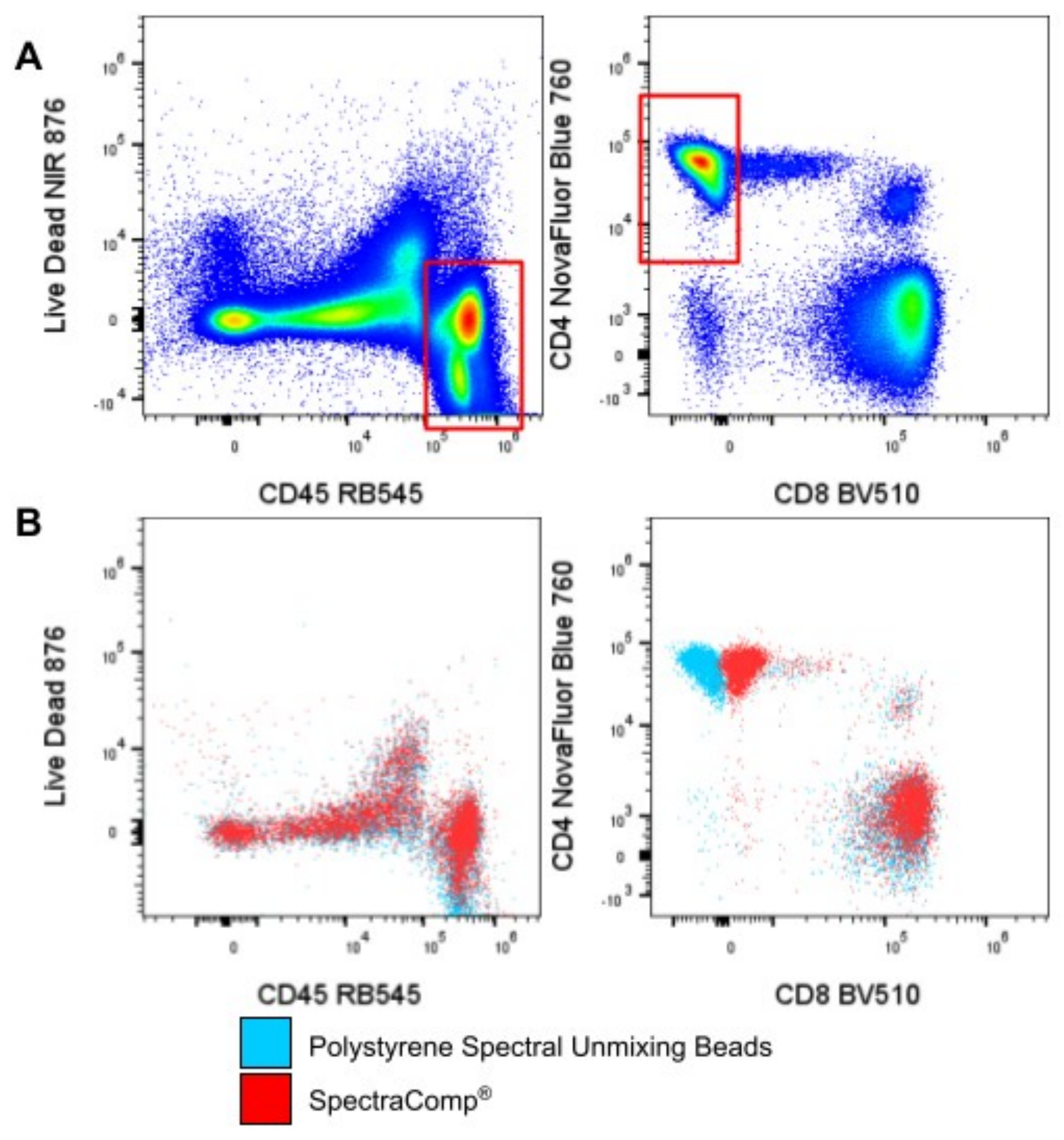
To address the unmixing errors, the BCM core chose to substitute 15 out of the 36 reference controls with SpectraComp® and ViaComp® cell mimic controls. After re-acquisition and unmixing, the problematic fluorophore combinations showed fewer unmixing errors with these new particles (Figure 1). This suggests that the cell-like properties of Slingshot cell mimics are beneficial for unmixing complex panels.
However, targeted substitution of the problematic fluorophores means a ‘mix and match’ approach with different control particles and does not facilitate a streamlined workflow. Using one type of control particle for the entire set of reference controls would make this panel an easy-to-implement tool for the BCM core to evaluate multiple instruments.
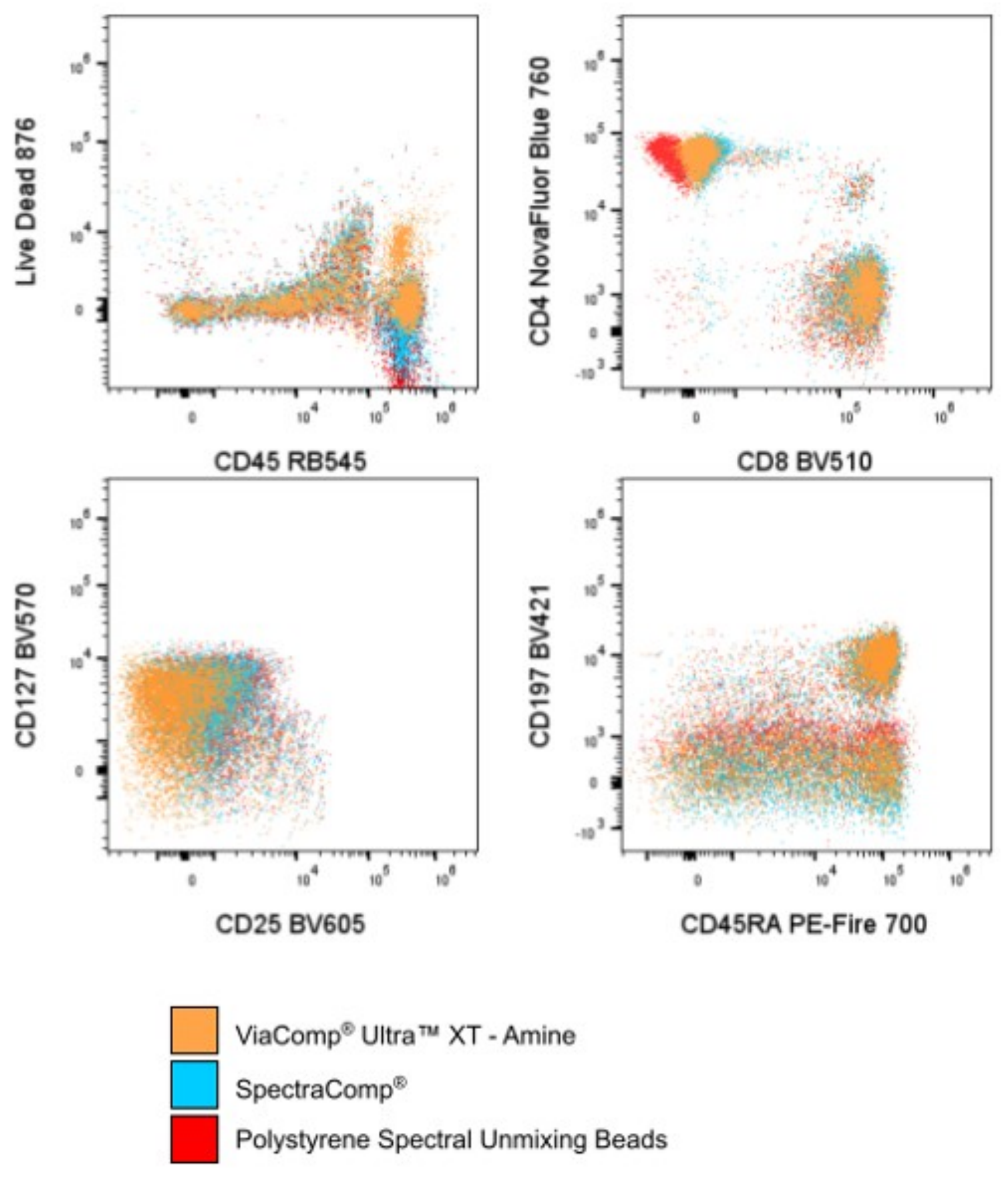
To streamline the workflow for BCM staff, all reference controls were replaced with ViaComp Ultra XT - Amine compensation particles. New single color antibody and fixable viability dye reference controls were acquired using ViaComp® Ultra™ XT – Amine as the control particle. The results resolved the original unmixing issues while enabling the desired streamlined workflow for implementation in the core (Figure 2).
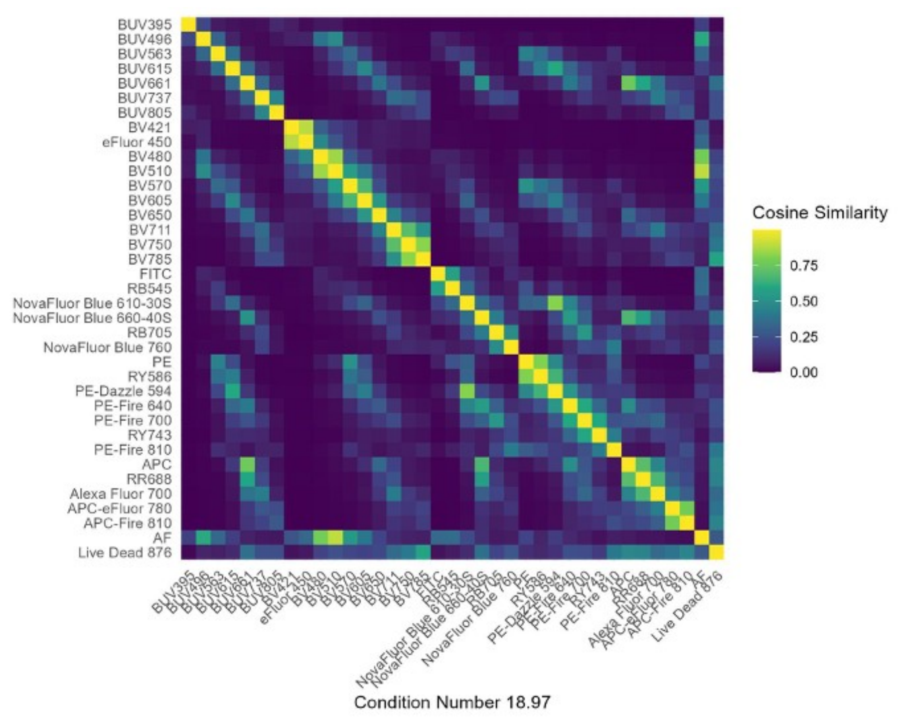
ViaComp® Ultra™ XT – Amine also resulted in reduced complexity of the similarity matrix of the complete panel. The overall degree of similarity of a spectral flow cytometry panel, referred to as the complexity index (condition number), is an indication of the overall difficulty of unmixing that combination of fluorophores.³,⁶ The similarity matrices were calculated based on the reference control spectra for each set of controls: polystyrene spectral unmixing beads, SpectraComp®/ViaComp® and ViaComp® Ultra™ XT – Amine. ViaComp® Ultra™ XT – Amine demonstrated a reduction in the complexity index of the entire panel, which resulted in more accurate unmixing of the entire panel (Figure 3).
| Control type | Complexity index |
|---|---|
| Polystyrene Spectral Unmixing Beads | 22.98 |
| SpectraComp®/ViaComp® Controls | 23.25 |
| ViaComp® Ultra™ XT – Amine Controls | 18.97 |
Table 2. Complexity index (condition number) by reference control type across the full 36-marker panel.
Conclusion
The use of accurate reference controls for spectral unmixing is critical to having reliable data from any spectral flow cytometry antibody panel. In developing and testing polystyrene unmixing beads, SpectraComp®, ViaComp® and ViaComp® Ultra™ XT – Amine controls, the latter proved to facilitate the best unmixing workflow for the development of this antibody panel. Use of ViaComp® Ultra™ XT – Amine resulted in decreased complexity, more accurate unmixing and facilitated a streamlined workflow for the implementation of this panel in the BCM core.
References
- Pilkington, K. R. Autofluorescence: From burden to benefit. Cytometry A 105, 563–567 (2024).
- Novo, D. A comparison of spectral unmixing to conventional compensation for the calculation of fluorochrome abundances from flow cytometric data. Cytometry A 101, 885–891 (2022).
- Park, L. M., Lannigan, J. & Jaimes, M. C. OMIP-069: Forty-Color Full Spectrum Flow Cytometry Panel for Deep Immunophenotyping of Major Cell Subsets in Human Peripheral Blood. Cytometry A 97, 1044–1051 (2020).
- Roederer, M. Compensation in Flow Cytometry. Curr. Protoc. Cytom. 22, (2002).
- Waaijer, L. A., van Cranenbroek, B. & Koenen, H. J. P. M. OMIP-112: 42-Parameter (40-Color) Spectral Flow Cytometry Panel for Comprehensive Immunophenotyping of Human Peripheral Blood Leukocytes. Cytometry A 107, 226–232 (2025).
- Mage, P. L., Konecny, A. J. & Mair, F. Measurement and Prediction of Unmixing-Dependent Spreading due to Collinearity in Spectral Flow Cytometry. Cytometry A (2026) doi:10.1002/cyto.a.70044.
Featured product
ViaComp® Ultra™ XT - Amine
slingshotbio.com/products/viacomp-ultra-amineSSB-35-B
 View all product info
For Research Use Only. Not for use in diagnostic or therapeutic procedures.
Stay Updated
Subscribe to receive product updates, new resources, webinars, and application notes.
Get the full application note
Download the complete note as a PDF, with the figures and reference, to share with your team.
Download PDF